

A Simulator on the Scene
August 07, 2024
Cardiology fellows, sonographers can practice testing with new echocardiogram
The cardiology fellow had just begun his rotation in performing and reading echocardiograms at University Hospitals Cleveland Medical Center when a new simulator arrived.
Now Tasveer Khawaja, MD, won’t only learn on live cardiac patients. He can step into the room housing the new echo simulator and practice with the latest 3D technology. The $92,000 investment by University Hospitals Harrington Heart & Vascular Institute is available 24/7 to its many cardiology fellows and cardiac sonographers training to perform transthoracic and transesophageal echocardiograms.
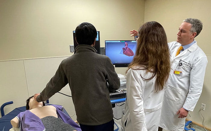
“One of the biggest challenges of learning transesophageal echocardiography is understanding the relative relationships of anatomical structures in 3D,” says Dr. Khawaja. “The simulator allows you to see echocardiogram-derived images and gross anatomy side-by-side. This is a huge benefit and has helped me understand imaging planes and how to optimize the TEEs I perform.”
Echocardiograms can aid in diagnosing a wide range of conditions, including mitral regurgitation, aortic stenosis, cardiac tamponade and endocarditis. The simulator contains all possible pathologies and explains diagnostics, so the user can read through the results even if an experienced physician isn’t at their side.
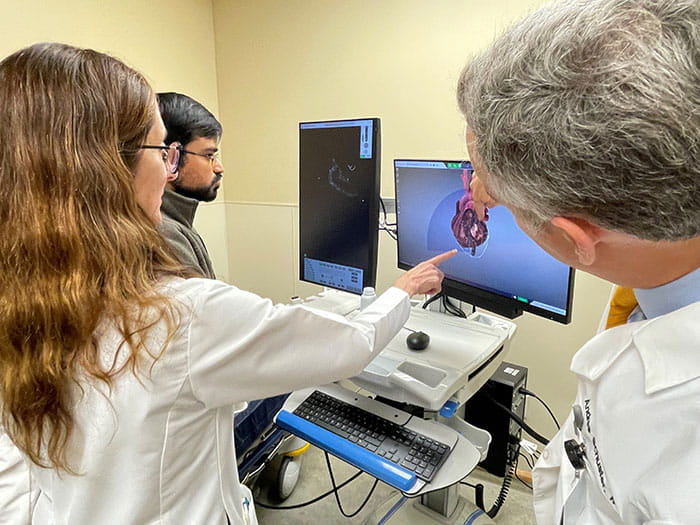
“You select the diagnosis you want to assess, and the simulator gives you a live feed, so you know if you’re finding what is expected,” says Andres Schuster, MD, the system medical director of adult echocardiography at UH Harrington Heart & Vascular Institute who was recently selected as Teacher of the Year by the cardiology fellows. “This is a really cool technology. I wish it was available when I was learning echocardiography.”
Senior staff cardiologist Ellen Sabik, MD, demonstrates for Dr. Khawaja how the images reveal the structure of the heart, allowing the user to slice through the heart and see 3D images. The echo equipment also assesses direction and speed of blood flow through the heart to determine whether it is functioning properly. The simulator allows the user to record a session for later review.
“We can learn a lot about physiology through this noninvasive testing,” Dr. Sabik says. “And we use echocardiograms to guide procedures like TAVR and MitraClip. We can use this simulation technology to learn how to better guide the interventionalists in doing these various procedures.”
Tags: Heart disease, Echocardiography