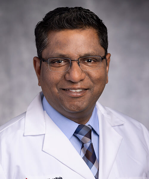
Introducing UH Rainbow's New Heart Expert | Arpit Agarwal, MD
November 12, 2023

Pediatric Clinical Update | November 2023
Dr. Agarwal joined the UH Rainbow Congenital Heart Collaborative in July 2023 following a national search and recently took a few moments for our brief interview
 ]Arpit Agarwal, MD
Arpit Agarwal, MD, FAAP, FACC, FASE, FSCMR, MS
Rainbow Chief Medical Information Officer
Pediatric Cardiologist with Advanced Imaging Expertise
Q: As chief medical information officer (CMIO), how has big data captured your passion? How will you use it to answer questions that ultimately benefit UH Rainbow patients?
As CMIO at UH Rainbow Babies & Children’s, I will strive to improve patient care by combining the best practices of pediatrics, computer science, information science and healthcare management. In this role, I am responsible for the design, implementation and use of healthcare information systems and technologies. It includes harnessing the information in electronic medical records to conduct data analysis and employ artificial intelligence to optimize operations, improve patient care, safety, and health outcomes. As CMIO, I work in six main domains:
- Clinical informatics
- Research informatics
- Operations, Finance and Policy informatics
- Quality informatics
- Public Health Informatics
- Consumer Health Informatics
In all these areas, I will work closely to support UH Rainbow administrators, physicians, and staff in utilizing data for informed decision making.
Q: You also have expertise in advanced imaging. How will you utilize these skills to help physicians, surgeons and patients in UH Rainbow Heart Center and throughout the hospital?
To answer the question fully, let me give some perspective. It was exciting for me to see how, in the latter half of the 19th century, advances in aerospace technology enabled scientists to send, maintain and repair spacecrafts, shuttles and rovers on the Moon and Mars while sitting millions of miles away on the Earth. But here on earth, we still are not able to make a complete diagnosis non-invasively and must get inside the heart to diagnose different ailments of the human heart.
This digital divide between medical science and information technology always intrigued me. I see myself as being able to help reduce this divide by using advanced three-dimensional (3D) imaging modalities, that we have had access to for the last two decades – such as CT, MRI, 3D echocardiograms. Now we are able to fully explore the potential of these 3D imaging modalities, by using advanced visualization technologies like, 3D printing, virtual reality (VR) and augmented reality (AR), opening a new paradigm of diagnostic imaging.
This cutting-edge technology helps specialists by providing them with the clearest possible picture of a patient’s complex anatomy. With 3D models they can attain a haptic experience and plan surgeries and other interventions, well in advance of the actual procedures. These 3D images and 3D printed models serve to help surgeons to create a customized surgical approach best suited for the individual patient. Having this information can also help reduce OR time, improve quality of care, patient safety, enhance patient and family education and ultimately improve outcomes.
Q: You are new to Cleveland – can you tell us about your training and career trajectory to date?
I earned my medical degree from King George’s Medical University in Lucknow, India, and completed a pediatric residency at Maimonides Infants and Children’s Hospital in Brooklyn, NY, followed by a pediatric cardiology fellowship at Jackson Memorial Hospital in Miami and a fellowship in advanced cardiac non-invasive imaging at University of Texas Health Science Center in Houston. Because of my interest in healthcare informatics, I went on to obtain a Master of Science degree in computer information systems from Boston University and completed the executive program in artificial intelligence in healthcare from the Massachusetts Institute of Technology. I am board certified in pediatrics, pediatric cardiology, cardiac magnetic resonance imaging (MRI) and clinical informatics. Prior to my current appointment at UH Rainbow, I served as chief medical information officer and director of advanced imaging at the Children’s Hospital of San Antonio and assistant professor at Baylor College of Medicine.
Q: What drew you to join The Congenital Heart Collaborative team at UH Rainbow Babies & Children’s Hospital?
I am passionate about building programs and am very excited to build a best-in-class clinical informatics program with state-of-the-art advanced imaging at UH Rainbow Babies & Children’s. After my first meeting with UH Rainbow President Patti DePompei and pediatrician-in-chief Dr. Marlene Miller, it was evident that we share this same passion, drive and vision for bringing the clinical informatics program and center of excellence for advanced imaging to life at UH Rainbow. I am also thrilled to be at UH Rainbow because of the legacy and rich history the hospital has among children’s hospitals.
Q: Can you tell us about your imaging background and how you plan to use this insight in treating patients at UH Rainbow? What is your plan for the next year?
In addition to my board certifications in pediatrics, pediatric cardiology and cardiac MRI, I am also board-certified in clinical informatics from the American Board of Preventive Medicine. I feel fortunate to have this unique combination of training which is shared by only handful of physicians in the country. My first goal is to map out and build a clinical informatics program at UH Rainbow. This will include establishing an informatics hub supported by a team of analysts to churn the data from EPIC and other data sources to answer critical questions regarding quality of care, patient safety, finance and healthcare operations. For example, when the hospital is looking to open a new facility, we can use data to determine the medical needs of the community, if the population in that community can support it, how many staff will be needed in each specialty, and likewise. The hub can also guide questions for research studies, disaster preparedness, financial feasibility and many other areas.
Concurrently, the next goal is to establish a Center of Excellence in Advanced Imaging that will encompass cardiology but also extend to orthopedics, plastic surgery, dental and many other specialties. My vision includes adding a state of the art cardiac CT, cardiac MRI, fetal MRI and 3D echocardiogram, and the purchase of equipment to lay the infrastructure for advanced visualization and 3D printing capabilities.
Once this foundation is in place, the imaging program will help surgeons who treat children with complex conditions. In these cases, the center will create 3D models ahead of the surgeries, so the specialists are equipped with as much detail about their patient’s condition and unique anatomy as possible. This helps them better plan and prepare for the procedures, resulting in shorter operative time and less complications. Patients will also benefit since the 3D models help clinicians educate patients and families about their child’s disease and how they plan to operate to correct the condition. In my prior institution, we gave patients a 3D replica of their heart, which helped them understand how it was repaired. It can become a souvenir -- but also the best education tool possible.
Q: What excites you about the expanding field of advanced imaging and computer vision within pediatric cardiology?
The most exciting part of advanced imaging is being able to diagnose the most complex diseases and help interventionists to improve patient outcomes. The field is ever evolving and there are endless possibilities. Right now, augmented reality (AR) is fast overtaking virtual reality (VR) as a preferred 3D visualization modality. VR limits physicians due to the complete immersion into the VR world. AR or mixed reality in contrast allows physicians to tap into 3D models while remaining grounded in the real world. Computer vision and AR takes the application of diagnostic imaging to the next stage of real time guidance to the surgeon in the OR. For example, AR allows you to understand the complex heart defect;
practice approaches that will be used in the operating room (OR) and then when surgery day comes, you can reference the 3D model as a guide throughout the case in real time.
Q: Can you share your research and education goals and interests?
Once we establish the clinical informatics program, our goal is to eventually build a strong fellowship in clinical informatics. Also, once the center for advanced imaging and 3D printing capabilities are up and running, we plan to start a fourth year fellowship in cardiac advanced imaging. I anticipate that an outgrowth of the fellowship program will include an explosion of research studies in pediatric cardiology and many other areas.
Q: As a relatively new Clevelander, have you explored the city much?
Right now I have been starting my day very early and leaving the hospital quite late. I found a home within walking distance of the hospital and have enjoyed these commutes. I find Cleveland to be quite beautiful and I am looking forward to exploring more of the city’s great restaurants, parks and culture.


