Advances in Diagnosis and Treatment of Lung Cancer
November 11, 2020
Updated screening guidelines could improve early lung cancer diagnosis, while new treatment options offer potential for patients with lung nodules
Innovations in Pulmonology & Sleep Medicine | Fall 2020
The U.S. Preventive Services Task Force (USPSTF) is updating its 2013 lung cancer screening recommendations, lowering the age for onset of screening to 50 and including more smokers.
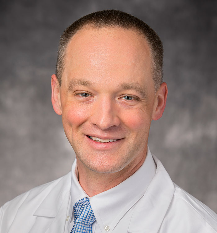
 Benjamin Young, MD
Results from the National Lung Screening Trial found that annual screening of people at high risk for lung cancer with low-dose computed tomography (LDCT) lowered their risk of dying from lung cancer by 15 to 20 percent. The USPSTF subsequently recommended lung cancer screening for individuals 55 to 80 years old who have a 30-pack-a-year smoking history and who currently still smoke or have quit within the previous 15 years.
“The new guidelines open up the benefits of lung cancer screening and early diagnosis to a larger, at-risk population,” says Benjamin Young, MD, Division of Pulmonary, Critical Care and Sleep Medicine at University Hospitals Cleveland Medical Center and Assistant Professor at Case Western Reserve University School of Medicine.
“Initial studies found a benefit to screening in very heavy smokers at high risk for lung cancer,” Dr. Young says. “However, in practice, we see that even people who smoked less are still at significant risk. The 55 age cut off was hindering many of them from getting screened.”
Lung cancer is the second most common cancer and a leading cause of cancer-related mortality. Furthermore, smoking accounts for roughly 90 percent of all lung cancer cases.
“The updated guidelines open screening to younger, heavy smokers and those who did not meet the 30-pack year threshold but are still at significant risk,” Dr. Young says. “Primary care physicians can now refer more patients with a history of smoking for lung cancer screening. Hopefully this will result in a stage shift away from patients with advanced lung cancer to patients with early stage lung cancer that is more readily treatable. We are also adding a robust database to our lung cancer screening program to better manage and care for enrolled patients.”
NEW TECHNOLOGIES FOR DIAGNOSING AND TREATING LUNG NODULES
UH is investing in the necessary equipment and technology to participate in several important clinical trials evaluating the diagnosis and treatment of pulmonary nodules.
Microwave thermal ablation. Although surgical resection is the gold standard for treating lung cancer, not all patients can undergo surgery. Microwave thermal ablation, can be performed both via a CT-guided percutaneous probe or a bronchoscopically placed catheter. CT-guided percutaneous ablation is a well-accepted technique, while bronchoscopic catheter ablation is a new therapeutic option currently undergoing clinical trials. Microwave ablation of pulmonary tumors may be an option for patients who have stage one lung cancer, but are not surgical candidates, and for those who have four or fewer metastatic nodules, no evidence of distant metastases, and a locally controlled primary tumor.
Direct injection of cytotoxic agents. Endobronchial Intratumoral Chemotherapy (EITC) provides local delivery of cytotoxic drugs to cancerous lung lesions and may provide an effective way to deliver chemotherapy (and other cancer-treating agents, such as immunotherapy) directly to a tumor, allowing clinicians to deliver higher local doses of drugs with fewer side effects.
Robotic bronchoscopy. Robotic bronchoscopy is FDA approved and has been in clinical practice since June of 2018. This technology allows clinicians to more accurately sample pulmonary lesions in the peripheral lungs, where approximately 80 percent of screen-identified lesions are located. Robotic bronchoscopy minimizes some of the limitations in existing bronchoscopy technologies. “Robotic bronchoscopy overcomes the limitations of current guided bronchoscopic techniques and improves the diagnostic yield on peripheral lung nodules,” says Dr. Young, an interventional pulmonologist who is Medical Director of Bronchoscopy at UH Cleveland Medical Center. “Improved diagnosis leads to earlier treatment for malignant lesions and peace of mind for patients with benign lesions.”
Machine learning and Radiomics1. Although not yet in clinical practice, the future of lung nodule diagnosis may include applying machine learning technology and radiomics (computer-extracted imaging features) to help clinicians predict whether nodules are malignant or benign based on criteria such as shape, texture and growth rate. Anant Madabushi, PhD, CWRU Professor of Biomedical Engineering, leads a team evaluating quantitative imaging analysis and signal processing for applications related to lung nodules and lung cancer in collaboration with Dr. Robert Gilkeson, Chief of Cardiothoracic Imaging at UHCMC, Dr. Philip Linden, Chief of Thoracic Surgery, and Dr. Frank Jacono, Chief of Pulmonary, Critical Care and Sleep Medicine. “This technology may help us risk-stratify lung nodules while preventing the need for invasive diagnostic procedures,” says Dr. Young.
MULTI-DISCIPLINARY EXPERTISE DRIVES COMPREHENSIVE CARE
Dr. Young notes that UH has a system-wide network of specialists in pulmonary medicine and thoracic surgery. Primary care and other referring physicians should know that their patients will be promptly and carefully evaluated and treated when necessary. In cases of lung cancer, UH pulmonology clinicians work with specialists at the UH Seidman Cancer Center.
For more information or to discuss a specific patient, call Dr. Young at 216-844-3201.
1J Med Imaging (Bellingham). 2018 Apr; 5(2): 024501, Radiology. March 2019; 290(3): 783–792.


