UH Seidman Cancer Center Early Adopter of New Ablation Technology for Liver Tumors
November 03, 2019
Advanced system provides patients with less invasive treatment options
Innovations in Cancer | Fall 2019
Surgeons at UH Seidman Cancer Center were among the first in the country to use a next-generation ablation system for treating primary liver tumors and liver metastases. After about a year with the new system, they say it’s making a measurable difference for their patients, allowing some access to laparoscopic procedures rather than open surgery.
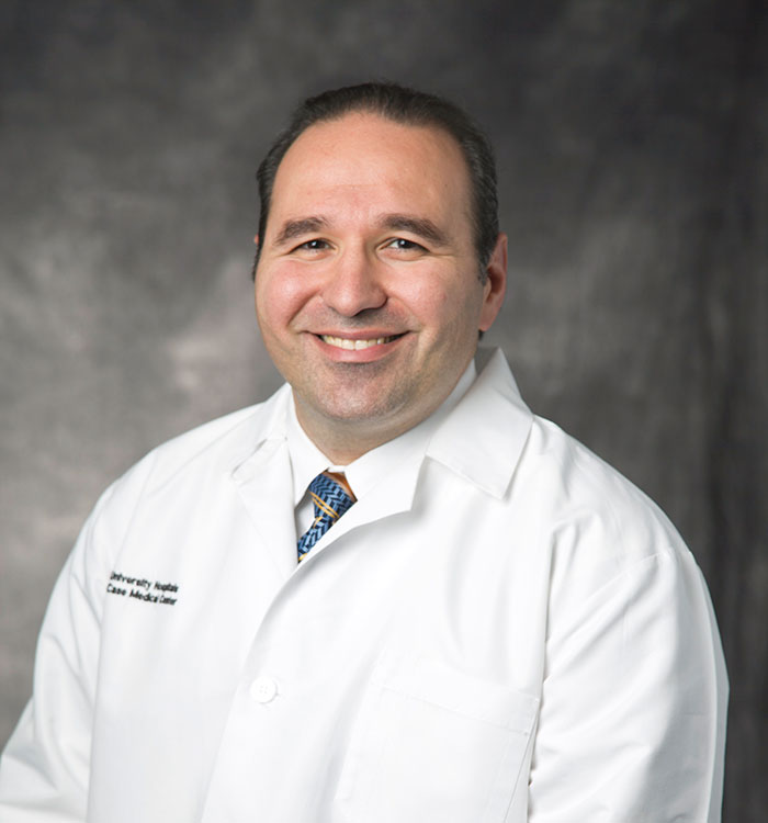
 John Ammori, MD
UH Seidman Cancer Center surgeon John Ammori, MD; and Associate Professor of Surgery, Case Western Reserve University School of Medicine uses the new system with his liver cancer patients. Here’s how he describes how it improves over older technology:
“For very small tumors or tumors in a difficult location, precise needle placement into the tumor with ultrasound guidance can still be very difficult,” he says. “You sometimes need to make multiple needle passes into the liver before finally hitting the target. Also, if the initial needle pass to ablate a small tumor is close but not perfect, the ultrasound picture may have some artifact from the initial needle pass, making it more difficult to hit your target.”
The new technology has made life a lot easier in terms of accuracy and the number of attempts to place the needle, Dr. Ammori says.
“It includes a board placed under the patient and a sensor on the ultrasound. The needle that we use for the microwave ablation also has a sensor in it. I place the ultrasound on the tumor and see the tumor deep within the liver. The sensing technology helps me direct the trajectory of the needle so that I precisely and accurately hit the target with one needle pass. I can manipulate the needle before I ever penetrate the liver so that I know I’m exactly on target. I can be 100 percent confident that I’m going to be right into the tumor.”
According to Dr. Ammori, the improved accuracy of the new ablation system is opening up new treatment possibilities for patients with liver tumors, whether primary or metastatic.
“Because of this, we’re sometimes able to do a procedure laparoscopically instead of open,” he says. “It just provides better accuracy. We usually observe people overnight to make sure there are no issues of bleeding. But it certainly takes people from a procedure that would require a few days in the hospital with a large incision, and it can turn it into an overnight procedure with minimal complications.”

The best outcomes for image-guided ablation among liver cancer patients are when the tumors are 3 centimeters or less or not located next to the central bile duct, Dr. Ammori says. It can be particularly well-suited for patients with hepatocellular carcinoma.
“Patients who have hepatocellular carcinoma often have cirrhosis and an abnormal liver,” he says. “This is a treatment that, rather than removing a large part of the liver, we can burn the tumor and little bit of normal liver around that tumor. It’s much easier and safer for the liver, with less risk for liver dysfunction or failure.”
Another common application of image-guided liver ablation is with patients who have metastases from either colorectal cancer or from neuroendocrine tumor, Dr. Ammori says.
“If you have patients who have multiple tumors that you can’t remove all the tumors by resecting them, there’s the opportunity for ablating some or all of the tumors and then resecting the other ones,” he says. “You can use it as a stand-alone treatment for all of the tumors in the liver, or you can use it in combination with resection. The goal is to kill or remove all of the liver tumors.”
Tumors deeply located in the liver are ideal candidates for image-guided ablation.
“Often deeply located tumors will require a major liver resection, sometimes even having to remove half or more of the liver because of the location of the tumor,” Dr. Ammori says. “However, if the tumor is deeply located and is of appropriate size for an ablation, you can ablate that tumor and significantly reduce the risk of treating that tumor. Essentially, it’s just as effective as far as the tumor outcomes go.”
For more information about image-guided ablation of liver tumors at UH Seidman Cancer Center, please call 216-553-1240.


