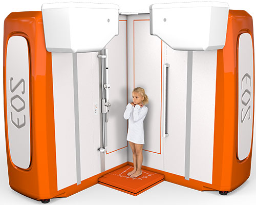
What is EOS Imaging?
EOS imaging is an X-ray technology that provides information-rich, weight-bearing images by scanning a patient in either a standing or sitting position using a low dose of radiation. The technology simultaneously captures frontal and lateral full body images in under 20 seconds for average-sized adults and 15 seconds for children.
Why Use EOS Imaging?
Many people, especially parents of young children and teenagers, are concerned about radiation exposure associated with X-rays or CT scans. Since EOS imaging uses lower doses of radiation than other imaging methods, it can be an appropriate alternative for children and adult patients being treated for conditions that require ongoing or frequent imaging.
What Is EOS Imaging Used For?

EOS Imaging can simultaneously take full-body, frontal and lateral images of the musculoskeletal skeletal system, making it useful in the diagnosis and treatment of medical conditions that affect the bones in the back, hip and leg. The technique is used for evaluating the following conditions:
- Balance and posture complications
- Degenerative disc disease
- Hip dysplasia in adults
- Hip dysplasia in children
- Kyphosis in adults
- Kyphosis in children
- Limb length discrepancy
- Osteoarthritis impacting the hip and knee
- Scoliosis in adults
- Scoliosis in children
- Bowleg and knock knee conditions
- Clubfoot and flatfoot
- Legg-Calvé-Perthes disease
- Slipped capital femoral epiphysis (SCFE)
- Patellar instability and patellofemoral pain
EOS Scan for Orthopedic Presurgical Planning and Postsurgical Assessment
Additionally, orthopedic surgeons can utilize EOS imaging in their presurgical planning, as the technology records and displays the patient’s anatomical structures in their true size, volume, lengths and angles. EOS imaging captures 1:1 functional 2D/3D images in under 20 seconds to support well-informed diagnoses, highly precise presurgical planning and postsurgical assessment for spinal surgery, hip replacement, knee replacement and other surgeries. For example, in spinal surgery, EOS imaging is used to:
- Generate 3D images of a patient’s spine in current and optimal corrected states
- Modify 3D spinal correction and implant positioning in real-time
- Export data to rod benders or a 3D printer
- Gain immediate understanding of 3D frontal and sagittal alignment
- Enhance surgical strategy by calculating post-operative parameters
Where Does UH Offer EOS Imaging?
We offer EOS imaging on the fifth floor of UH Cleveland Medical Center Bolwell.
UH Cleveland Medical Center – Bolwell Building, 5th floor
11100 Euclid Avenue
Room 1259
Cleveland, OH 44106
What to Expect Before and During an EOS X-Ray
Patients can eat normally before having an EOS scan . Certain types of clothing and jewelry can interfere with the images and will have to be removed prior to the exam. Patients may need to change into a gown or paper shorts for their EOS X-ray depending on the part of the body that needs imaging. Because EOS imaging utilizes a very low dose of radiation, patients do not have to wear a protective lead apron.
During an EOS exam, the patient stands still or sits upright inside a scanning cabin. Two very narrow X-ray beams – one vertical, one horizontal – scan the entire body to create 2D and 3D images of the spine and joints. Unlike conventional X-ray imaging, where the patient may have to be repositioned to capture images from different angles, these two simultaneous scans provide all the imaging necessary.
What Happens After an EOS X-Ray?
After an EOS exam, a specially trained radiologist interprets the X-ray images and sends the results to the doctor who ordered the exam. If the results are urgent, the radiologist will contact the doctor immediately.
Do EOS X-Rays Cost More Than Traditional X-Rays?
2D EOS x-rays cost the same as traditional x-rays and are billed in the same manner. 3D EOS images are billed at an additional cost to the original 2D EOS scan. Coverage of the 3D portion varies from insurer to insurer.


